
A Producer’s Guide to Encountering Disease in Broilers
By Dr. Linnea Newman
Features New Technology ProductionEncountering Disease in Broilers
They’re Dying – 100 a day – What Do I Do Now?
Introduction
Few things are more disheartening to a poultry producer than to walk into a flock and find birds dying when they’d looked just fine the day before. It means a lot more work ahead and less profit at the end of the flock despite the effort.
Although poultry disease is the realm of a veterinarian, there are some basic diseases that producers can recognize on their own, and there are some basic things that they can do to give their veterinarian or health advisor a head start.
What follows is a troubleshooting guide for some of the common and emerging diseases and conditions that producers may encounter in future flocks.
Days 1 to 7: Chick Quality and Brooding Management
Tool: Post-mortem
How many? 25 chicks or more
Which ones? Dead chicks (not culls)
Age: 3-4 days only
It is nearly impossible to distinguish the cause of mortality in baby chicks without postmortem of the dead chicks at about 3 to 4 days of age. It may be too early to see much in chicks younger than 3 days, and too late to distinguish the source of a problem in chicks older than 5 days. Freshly killed chicks, even sick ones, rarely provide a clear answer.
Where mortality is a problem, it will be easy to find 25 dead chicks or more. There will be a mixture of problems in a large population of birds, so just make piles of birds for each category: the largest pile is the root of the high mortality problem. The focus will be on the navel, the yolk, the lungs and the kidney.
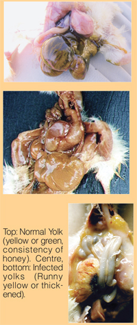
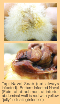 Conditions Causing High Chick Mortality:
There are only three major causes of high mortality in baby chicks. They are easy to distinguish. Learn to watch out for number 3 – dehydration – so that corrective management action can taken. The producer has the greatest influence on dehydration.
1. Omphalitis: Omphalitis is characterized by nasty-smelling yellow stuff in the belly.
a. Yolk sac infection
This is an infection involving the entire yolk. The yolk is thin and runny – or thick like cottage cheese. This indicates an early bacterial contamination of the egg due to dirty eggs, wet eggs or thin shells … and the source is often the breeder flock rather than the hatchery. Older flock sources are particularly prone to producing chicks with yolk sac infection because the pores of the egg are naturally larger and easier for bacteria to penetrate.
Treatment: cull.
b. Navel infection
An infection originating from the navel, but the yolk is normal. This may be difficult to distinguish from a yolk sac infection, especially if it is severe. Navel infections are more likely to be hatchery-origin.
Treatment: cull.
Treatment: The day-of-age antibiotic given to chicks at the hatchery was the strongest treatment they could receive. Those that succumb to infection anyway should be culled. Exception: low-grade navel infection may result in E. coli during the second week. Such flocks may benefit from water antibiotic treatment (see E. coli in the next section).
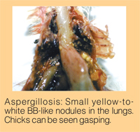 Aspergillosis
A mold infection characterized by little tiny yellow or white nodules or “BB’s” in the lungs and upper airsacs. This infection is often due to contamination of incoming eggs, or of the ventilation system in the hatchery but can also come from new wood shaving litter. Mortality can already be high at three days of age from a litter-origin infection.
Treatment: cull.
Starve-out / Dehydration
This problem is characterized by small, weak chicks. It is most common in chicks from young breeder flocks placed in the winter. The number one contributor to this problem is brooding management. Chicks chilled by cool or damp floors tend to be sluggish and never move properly to feed and water.
Poor water access due to water pressure, drinker height or air lock problems can also quickly produce dehydration in young chicks. Beware of thin litter, lack of radiant brooders, floor condensation and low floor temperatures: all are common contributors to this problem.
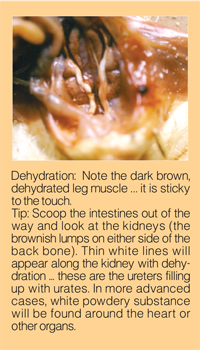 Treatment: cull. Walk the chicks to encourage movement to feed and water while management problems are being corrected.
Day 7 and Beyond: After the First Week
Tool: post-mortem
How many? 8 birds
Which ones?
2 random, healthy birds
2 smaller, apparently healthy birds
2 definitely sick or down birds
2 dead birds
Tip: Take the time to become familiar with “normal.” Use the random, healthy birds for comparison.
Day 7 to 21 – Conditions Causing High Mortality
E. coli “Creeping crud”
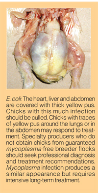
Low-grade E. coli infection from the navel can balloon into massive systemic infection during the second or third week, especially if flocks are stressed (brooding) or if they’re also exposed to an immunosuppressive problem like inclusion body hepatitis (see below). Yellow cheesy stuff will envelope the heart, the liver and fill the body.
Treatment: Use your bird sample as a guide. If the random and smaller birds are clean, you may be able to clear up the flock with culling alone.
If the smaller “healthy” birds, or even the larger random birds show signs of infection, antibiotic treatment with a broad-spectrum water soluble product (tetracycline, chlortetracycline, oxytetracycline, erythromycin) is warranted.
 Emerging Problem: Runting and Stunting Syndrome Day 6 to 10
This is an emerging U.S. problem that may be related to management and/or infectious causes. Flocks show a significant loss in uniformity evident by 10 days and develop signs of immunosuppression (gangrenous dermatitis and E. coli) later in life. Flocks have vent pasting, feed passage and thin-walled intestines with watery contents. Many birds have an enlarged proventriculus.
The problem is most pronounced in high-density, small bird or Cornish flocks and may have been intensified by short down time common in the U.S. this year.
Treatment: None so far. The U.S. is still trying to find out what it is. Cleaning, disinfection and increased down time appear to help.
Emerging Problem: Inclusion Body Hepatitis (IBH)
IBH is caused by an adenovirus that is transmitted from the breeders directly to the baby chicks. Signs of the virus transmitted directly from breeders to the chicks will appear at 10 -12 days of age. The virus can also spread from chick-to-chick or seed a broiler barn for future flocks. Chick-to-chick infection tends to produce signs at 16 to 17 days, up to about 22-35 days of age. The problem is characterized by a massive mortality spike. Sick and dead birds may have enlarged livers that may be greenish, yellowish or bloodshot. Intestines are normal.
Treatment:
a. Minimize the opportunity for simultaneous immunosuppression due to
infectious bursal disease (IBD) by IBD vaccination of broilers at day-of-age and in the field. Pay particular attention to the spectrum of protection to include protection against variant IBD strains.
b. Treat secondary E. coli infection based on your bird sample (see E. coli above).
c. A vaccine is currently under development for breeder pullets to reduce or eliminate virus shed to the broiler progeny. Until the vaccine is completed, pullet growers must consider the risky practice of spreading litter from exposed flocks into naive pullets to achieve some immunity.
Necrotic Enteritis (NE)
Like IBH, this problem causes a sudden, massive mortality spike. The cause is a bloom of Clostridium perfringens bacteria in the intestine. These bacteria (from the same family that produces botulism) produce a deadly toxin that leaks into the bloodstream. The bacteria bloom in conditions of high intestinal pH, and they are helped along by anything that irritates the intestinal lining (including wheat rations and coccidial infection). The problem will not be evident in any random birds, and may not even be obvious in sick birds. The dead birds will have swollen, ballooned intestines. The inside of the intestine is rough (normal intestine is fairly smooth) and often has the appearance of a brown turkish bath towel. The common times to see NE: flocks using a coccidiosis vaccine will experience NE at 16 to 17 days (the peak of vaccination reaction). Flocks using an in-feed ionophore coccidiosis control medication will usually experience NE later: 26 to 35 days (the peak of oocyst leakage with in-feed products). By contrast in-feed chemical coccidiosis control medication may have problems before day 12.
Treatment: Immediate treatment with penicillin, bacitracin or other antibiotic effective against Clostridium via drinking water.
Prevention:
a. Minimize wheat in the ration and use enzyme preparations to improve digestibility. Avoid wheat in the starter ration when possible.
b. Control litter moisture. High litter moisture increases the opportunity to develop NE.
c. Acidify drinking water for the week prior to the age of the typical break on a given program.
d. Salt dirt floor houses between flocks (top dress well with litter).
Chicken Infectious Anemia (Chicken Anemia Virus, Blue Wing) – Day 14 to 16
Chicken anemia, like IBH, is caused by a virus that is transferred from breeders exposed during production to the broiler progeny via the egg. Affected broilers look great until day 14, when they suddenly begin to cull up. Some of the broilers may develop gangrenous dermatitis, especially on the wings (“blue wing disease”). The blood becomes thin, like strawberry Kool Aid®, the bone marrow is pale and the thymus gland on the back of the neck becomes atrophied. Mortality will spike for a week, and the flock may return to normal growth or may develop secondary bacterial infections.
Treatment: Immediate treatment with penicillin, bacitracin or other antibiotic effective against Clostridium bacteria (the cause of dermatitis as well as necrotic enteritis). Water soluble vitamin E may also help recovery.
Prevention:
a. Vaccination of the pullets with CAV vaccine prior to production.
b. Avoid day-of-age IBD vaccination of progeny of shedding breeder flocks. The vaccine at day old can make the blue wing worse. Field boosting is ok.
Spiking Mortality (Hypoglycemia) + Rickets
“Spiking” says it: big healthy-looking birds suddenly begin to die for no apparent reason. Post-mortem shows absolutely nothing wrong with these dead or dying birds except a gizzard full of shavings. A blood test with human diabetic’s test strips (from the drug store) will show almost no blood sugar in the down, dying birds. Blood sugar will be normal in the random, healthy birds. Spiking mortality is caused by either a true feed outage or a “perceived” feed outage. It is very common with half-house brooding – occurring at 12 to 16 days of age when flocks are moved from half house to full house. Insufficient feed (caused by sudden removal of supplemental feeders or failure to trigger the control pan in the back of the house) can start the mortality event. Perceived lack of feed (houses too cool or too dark, or insufficient caloric content in feed) can also cause a spiking event. Birds that don’t actually spike may develop rickets (wide growth plates in the bone) instead.
Treatment: The spike will resolve as quickly as it started. Some have reduced the losses by placing feed on feed lids sprinkled with powdered milk to attract the birds. Water soluble vitamins, including A, D and E will aid the recovery and help to reduce the rickets that often follows.
Prevention: Care to avoid accidental feed restriction through temperature, lighting or feed management is critical. A lighting program between days 4 and 22 with dark periods ranging from 6 to 10 hours controls spiking mortality.
Days 21 to 35 – Conditions Causing High Mortality
Tools: Routine post-mortem of 5 random, healthy birds at 28 days.
In case of disease, follow the 8-bird rule: 2 healthy, 2 smaller healthy, 2 obviously sick, 2 dead.
Coccidiosis
Coccidiosis is caused by a single-cell intestinal parasite in the genus Eimeria. Most broiler mortality is due to a specific species: Eimeria tenella. E. tenella causes bloody droppings and can produce a sudden spike in mortality, usually between 21 and 35 days of age. Post-mortem of dead or down birds will show blood in the cecal pouches … random birds may not have any signs.
More importantly, if E. tenella is a problem, another species of coccidia may also be causing significant damage: E. maxima. E. maxima is often a silent infection; it may only cause mild ballooning in the middle intestine – even when severe. But where E. tenella kills, E. maxima destroys feed conversion.
The final common coccidial species, E. acervulina, forms white spots inside the intestine in the loop immediately after the gizzard. E. acervulina does little serious harm alone, but may be a warning that silent E. maxima is also at work.
Coccidiosis becomes a serious problem when the house coccidial population becomes resistant to feed preventatives or when coccidiosis vaccination is incomplete. If resistance occurs to many anticoccidial drugs, vaccination will achieve the dual purpose of immediate protection and reseeding the houses with anticoccidial-sensitive strains of coccidia.
Treatment: Amprolium at full treatment level. Follow with a course of water-soluble vitamins including A, D and E for more rapid recovery.
Prevention: Anticoccidial medication in the feed or coccidiosis vaccination at the hatchery. Routinely monitor flocks at 28 to 30 days to make sure coccidiosis is truly under control. Post-mortem five random birds per house at this age, every flock. Low-grade coccidiosis can silently rob performance.
Necrotic Enteritis
See the necrotic enteritis summary in the Day 7 to 21 section. NE may occur in this age range following moderate to severe coccidial damage.
Inclusion Body Hepatitis
See the IBH summary in the Day 7 to 21 section. IBH may occur in this age range from bird-to-bird transmission starting with a few “seed” chicks from positive breeder flocks.
Spiking Mortality
See the Spiking Mortality summary in the Day 7 to 21 section. Spiking mortality at 28 days and cellulitis are often related to the same management issues. See cellulitis below.
Airsacculitis / Respiratory Disease
Airsac disease is usually accompanied by coughing or sneezing or rales in the flock. Upon post-mortem, yellow suds or cheesy material may be seen in and around the organs or overlaying the pink lungs in the rib cage. Airsacculitis may indicate infection with a respiratory virus (Newcastle or bronchitis) or may result from the live vaccines given earlier in the flock’s life. Reaction to live respiratory vaccine is very often the cause of airsac disease in a young flock. Infectious bronchitis (especially variant strains) is often the cause in older flocks.
The yellow airsacculitis lesion is caused by E. coli, which we can treat with antibiotics. The root cause of the airsacculitis in commercial broilers is usually a virus, and a laboratory’s help will be needed to find the real culprit so that airsacculitis can be prevented in the future. Beware of Mycoplasma spp. infection in back yard birds or broiler chicks purchased from hatcheries that are not Mycoplasma free.
Treatment: The necessity of treatment is based on the severity of infection. Flocks with airsacculitis lesions in the random and smaller birds will require treatment with an effective broad-spectrum antibiotic (see E. coli). Flocks that only have airsacculitis lesions in down or dead birds may respond to culling alone. Note that high mortality due to airsacculitis almost always requires treatment, but treatment isn’t always successful.
Get help with diagnosis! Respiratory disease – no matter what age – could be avian influenza. Better to know than to be the source of a serious problem.
Prevention: Seek the advice of a health professional. Airsacculitis may be the result of rolling vaccination reaction, a new respiratory virus (including variant bronchitis or AI), or a result of immunosuppression. Airsacculitis is often induced by a key management stressor. We need a careful diagnosis before a prevention strategy can be implemented.
Cellulitis (Inflammatory Process, IP)
Ok, this doesn’t cause high mortality, but it can cause high condemnation at processing. Cellulitis is created by scratches that occur during the 21 to 35-day window of growth, when the birds’ flanks are feathered the least.
The same things that cause spiking mortality in younger birds can lead to cellulitis at the plant: feed restriction or perceived feed restriction. The result is too many birds trying to access the feed pans at one time. Most houses are equipped to allow birds to eat in four shifts. Disruption of these shifts by anything that stops the birds from eating for a period of time (lighting programs, cool or hot house temperatures, timed feeding, feed outage) can cause too many birds to jam the feeders at once. Anything that causes one shift to take too long to eat (low feed in pans, mash instead of pellets, low calorie feed) can also cause the birds “waiting in line” to get impatient. The end result: scratches and cellulitis.
Treatment: none
Prevention: Observe the birds carefully during feeding. Set the alarm for 1:00 am, or bring a good book with a reading light and a cup of coffee to spend a night with the birds at around 30 to 35 days of age. Watch what happens at sundown, at sunrise and when lights come on at night. The scratches often happen when no one’s watching. Watch for crowding, climbing on each other and tipping feed pans as birds fight for access to the feed.
Cholangiohepatitis “Hepatitis Condemnation”
Like cellulitis, this problem doesn’t kill, but causes heavy condemnation losses. Earlier Clostridium bacterial infections (see Necrotic Enteritis) may be the cause.
Emerging Problem: Variant Infectious Bursal Disease (IBD)
IBD generally doesn’t directly cause broiler mortality, either. But it does set the stage for mortality due to other diseases later on. Variant IBD causes rapid atrophy of the bursa at a young age (before 21 days). Bursas should be slightly smaller than the size of a quarter at about 28 days. Smaller bursas indicate exposure to IBD. As variant IBD spreads west across Canada, bursal atrophy is occurring earlier in spite of solid IBD vaccination programs.
Treatment: none.
Prevention: Keep an eye on bursas between 21 and 28 days of age. Post-mortem five random birds per house every flock between these ages. (A five-bird sample at 28 days will allow you to keep an eye on coccidiosis and the bursas.) If early atrophy is observed, consider switching to a vaccine that provides broader coverage against variant strains. A routine exam of five random, healthy birds at 28 days will give you an opportunity to monitor the health of the bursa and to watch out for low-grade coccidiosis that robs performance.
Day 35 to Processing – Conditions Causing High Mortality
Common Late Problem in the U.S.: Gangrenous Dermatitis
Gangrenous dermatitis is caused by Clostridium perfringens, the same toxin-producing bacteria that causes necrotic enteritis. Processing-aged birds die very quickly and look like they’ve been dead for hours, even though the bodies may still be warm. The skin often looks bruised and purple. Under the skin is a purple jelly that often contains gas bubbles. The liver may be enlarged and there may be some internal hemorrhaging. The disease is often found in flocks that have been immunosuppressed by IBD, CAV or IBH.
Treatment:
a. Antibiotics: penicillin, bacitracin or other antibiotic effective against Clostridium bacteria.
b. Frequent collection of mortality to avoid spreading due to birds picking at contaminated carcasses.
Prevention:
a. Protection of the immune system against immunosuppressive viruses through vaccination of the parent flocks or the broilers themselves.
b. Frequent collection of mortality to prevent picking at the dead carcasses.
Respiratory Disease
See “Airsacculitis/Respiratory Disease” in 21 to 35 day old flocks. It is very important to do a post-mortem on the suggested eight-bird sample whenever late mortality with respiratory noise is experienced. Respiratory disease may be broken into three critical groups based upon post-mortem observations:
Newcastle or Infectious Bronchitis (Including Variant IB)
These are the “common” respiratory viruses. They are usually followed by the typical secondary E coli airsacculitis (yellow suds or cheesy material in the body cavity and covering organs).
Treatment: Broad-spectrum antibiotics for the E. coli infection (see E. coli).
Prevention: Adjust vaccination program with the help of health professionals.
Laryngotracheitis Infection (ILT)
LT is not common, but has caused frequent outbreaks in Ontario. Severe respiratory noise is evident in the flock, and mortality is high. The eyes are often weepy or squinty. The only post-mortem lesion tends to be a very, very red inner trachea. Sometimes the trachea is filled with blood or yellow cheesy material. Often, dead birds will have the characteristic lesion while random and down birds do not. Typically, LT does not produce the typical yellow lesions of airsacculitis.
Treatment: Air and prayer.
Prevention: Vaccination. Prior to cleanout, litter may be composted in the house and house temperature should be raised to 100 degrees or more. LT virus is very heat-sensitive.
Avian Influenza (AI)
AI is tricky. Although the first incident is more likely to strike older flocks, it can affect flocks of any age! In AI’s worst form, airsacculitis may be present, but it will be in conjunction with lots of other problems: enlarged liver and spleen, blood-spotted intestines, blood-spotted internal gizzard or proventriculus … blood-spotted everything. AI makes the blood vessels leaky, so hemorrhage can be present anywhere. Even the legs may have purple bruising. In its mild forms, AI can easily be confused with plain old infectious bronchitis or Newcastle disease. This is why it’s important to seek the help of a health professional whenever respiratory disease is involved. AI can be tracked from flock to flock, appearing as a mild respiratory disease in summer weather, only to return to it’s ugly self when the weather turns cold.
Treatment: Air and prayer.
Prevention: Careful biosecurity!
Note: High mortality with respiratory signs is DANGEROUS. Do NOT call friends to come and see your birds in case they know what the problem is. Do NOT go visit another producer’s farm for advice and help. Get professional help before your farm becomes the index (original) case from which LT or AI spreads throughout the province.
Leg Problems
Leg problems can be a nagging source of culls, or they can be the cause of high mortality. While there are many potential causes of leg problems, two are the most common:
Tibial dyschondroplasia (TD)
TD is caused by a cartilage plug at the top of the tibia. Slice the leg bone lengthwise, just below the “knee.” TD makes the bone hard to slice. Once the cut is made, the entire top of the bone will be filled with a large, soft white plug of material. TD can have several causes, including mycotoxins, but is most often due to an imbalance of calcium and phosphorus in the ration, with phosphorus too high. Individual birds with TD may have experienced feed separation at the end of the feed line or may be genetically susceptible.
When an entire flock is severely affected with TD, have the feed tested. The problem may have been caused by earlier feeds, so always save samples of each feed during the flock.
Treatment: None. Cull the affected birds.
Prevention: Commercially formulated and manufactured rations. Call a health professional if repeat problems are
experienced.
Joint or Bone Infection
Joint infection is “synovitis” and bone infection is “osteomyelitis.” The key sign in each is yellow stuff. A normal leg joint should have just a tiny bit of clear or yellow-tinged fluid between the bones. Infection often affects the hock, but sometimes the knee joint, and is characterized by lots of thickened yellow fluid or pus in the space between the bones. Infection of the bone itself is characterized by yellowed areas of the interior bone. A lengthwise cut of the tibia will reveal a small spot of yellow within the normal pink-coloured bone near the top, just below the white or grey cartilage on the end. Leg infection is usually due to a bacterial infection (Staph or E. coli) that has gone systemic. The body’s immune system can fight the infection off everywhere that’s reached by the blood stream, but the joints have a minimal blood supply, so bacteria can safely hide and multiply.
The sources for the original bacterial infection can include:
Baby chick navel infection: Low-grade infection that has become chronic and eventually finds a home in the legs.
Enteritis (including coccidiosis, necrotic enteritis, or general gut irritation): Damage to the intestinal wall allows the normal intestinal bacteria to leak into the blood stream and become systemic.
Airsacculitis: Bacteria from respiratory disease will often find its way to the legs.
Immunosuppression (including viruses, heat stress or management stress): An impaired immune system can’t clear normal daily bacterial insults from the system fast enough to avoid some bacteria finding the legs. Viral immunosuppression (IBD, CAV, IBH) can result in heavy losses due to bacterial leg problems. Chronic or severe stress is also suppresses the immune system: high stocking density (chronic stress) or heat stress (chronic or severe) can result in leg problem losses. U.S. integrators have also induced leg problems with very low protein or cheaply formulated feeds.
Treatment: Treatment can be attempted with broad-spectrum antibiotics, but is rarely successful. Blood carries antibiotics to affected tissue, and there is very little blood supply in the joints. Culling is often the best option.
Prevention: Keep a close watch for signs of coccidiosis or IBD at the routine 28-day post-mortem session. Control stressors: avoid excessive stocking density and install cooling systems for summer heat.
Conclusion
No one expects a producer to be their own veterinarian, but producers can certainly learn to recognize some basic disease conditions. Armed with this knowledge, a producer can use their post-mortem information to make wise choices in treatment, to make corrections in management or to give useful information to health professionals for effective help.
Dr. Newman joined Schering-Plough Canada as their Technical Service Veterinarian in 2006. She can be contacted through your Schering-Plough representative.
Print this page